Kirjojen hintavertailu. Mukana 12 595 353 kirjaa ja 12 kauppaa.

Kirjailija

Igor Pronin

Kirjat ja teokset yhdessä paikassa: 6 kirjaa, julkaisuja vuosilta 2014-2019, suosituimpien joukossa Neuroimaging of Traumatic Brain Injury. Vertaile teosten hintoja ja tarkista saatavuus suomalaisista kirjakaupoista.

6 kirjaa

Kirjojen julkaisuhaarukka 2014-2019.

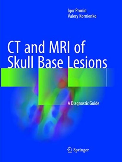
CT and MRI of Skull Base Lesions

CT and MRI of Skull Base Lesions

Igor Pronin; Valery Kornienko

Springer International Publishing AG
2019
nidottu
This superbly illustrated book offers a comprehensive analysis of the diagnostic capabilities of CT and MRI in the skull base region with the aim of equipping readers with the knowledge required for accurate, timely diagnosis. The authors’ vast experience in the diagnosis of skull base lesions means that they are ideally placed to realize this goal, with the book’s contents being based on more than 10,000 histologically verified cases of frequent, uncommon, and rare diseases and disorders. In order to facilitate use, chapters are organized according to anatomic region. Readers will find clear guidance on complex diagnostic issues and ample coverage of appearances on both standard CT and MRI methods and newer technologies, including especially CT perfusion, susceptibility- and diffusion-weighted MRI (SWI and DWI), and MR spectroscopy. The book will be an ideal reference manual for neuroradiologists, neurosurgeons, neurologists, neuro-ophthalmologists, neuro-otolaryngologists, craniofacial surgeons, general radiologists, medical students, and other specialists with an interest in the subject.
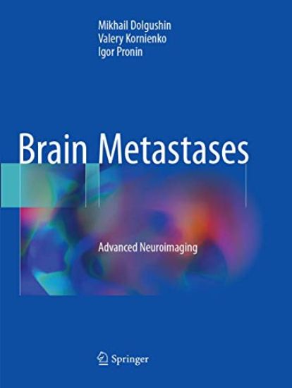
Brain Metastases

Brain Metastases

Mikhail Dolgushin; Valery Kornienko; Igor Pronin

Springer International Publishing AG
2018
nidottu
This book describes the role of advanced neuroimaging techniques in characterizing the changes in tissue structure in patients with brain metastases. On a large number of newly recognized CT, MRI, and PET characteristics of brain metastases from different primary tumors are highlighted, thereby elucidating the potential differential diagnostic role of CT perfusion imaging, MR spectroscopy, MR diffusion-weighted imaging, MR susceptibility-weighted imaging, and PET with different radiopharmaceuticals. For example, the different manifestations of metastases of melanoma, renal cell carcinoma, and ovarian cancer on MRI and CT perfusion imaging are described, and the role of MR susceptibility-weighted imaging in the differential diagnosis of glioblastoma multiforme and metastatic tumors is clarified. Metastases of colon cancer have shown a special manifestation on T2 weighted images. The book also presents novel findings regarding pathogenesis and tumor biology and describes qualitative and quantitative changes in tumor tissue and alterations in brain white matter due to surrounding tumor growth. Neuroradiologists and others, including neurosurgeons, neurologists, and nuclear medicine physicians, will find that this book offers a fascinating insight into the ways in which newly available data on structural, hemodynamic, and metabolic changes are enriching the neuroimaging of brain metastases.
CT and MRI of Skull Base Lesions

CT and MRI of Skull Base Lesions

Igor Pronin; Valery Kornienko

Springer International Publishing AG
2018
sidottu
This superbly illustrated book offers a comprehensive analysis of the diagnostic capabilities of CT and MRI in the skull base region with the aim of equipping readers with the knowledge required for accurate, timely diagnosis. The authors’ vast experience in the diagnosis of skull base lesions means that they are ideally placed to realize this goal, with the book’s contents being based on more than 10,000 histologically verified cases of frequent, uncommon, and rare diseases and disorders. In order to facilitate use, chapters are organized according to anatomic region. Readers will find clear guidance on complex diagnostic issues and ample coverage of appearances on both standard CT and MRI methods and newer technologies, including especially CT perfusion, susceptibility- and diffusion-weighted MRI (SWI and DWI), and MR spectroscopy. The book will be an ideal reference manual for neuroradiologists, neurosurgeons, neurologists, neuro-ophthalmologists, neuro-otolaryngologists, craniofacial surgeons, general radiologists, medical students, and other specialists with an interest in the subject.
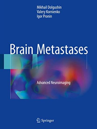
Brain Metastases

Brain Metastases

Mikhail Dolgushin; Valery Kornienko; Igor Pronin

Springer International Publishing AG
2017
sidottu
This book describes the role of advanced neuroimaging techniques in characterizing the changes in tissue structure in patients with brain metastases. On a large number of newly recognized CT, MRI, and PET characteristics of brain metastases from different primary tumors are highlighted, thereby elucidating the potential differential diagnostic role of CT perfusion imaging, MR spectroscopy, MR diffusion-weighted imaging, MR susceptibility-weighted imaging, and PET with different radiopharmaceuticals. For example, the different manifestations of metastases of melanoma, renal cell carcinoma, and ovarian cancer on MRI and CT perfusion imaging are described, and the role of MR susceptibility-weighted imaging in the differential diagnosis of glioblastoma multiforme and metastatic tumors is clarified. Metastases of colon cancer have shown a special manifestation on T2 weighted images. The book also presents novel findings regarding pathogenesis and tumor biology and describes qualitative and quantitative changes in tumor tissue and alterations in brain white matter due to surrounding tumor growth. Neuroradiologists and others, including neurosurgeons, neurologists, and nuclear medicine physicians, will find that this book offers a fascinating insight into the ways in which newly available data on structural, hemodynamic, and metabolic changes are enriching the neuroimaging of brain metastases.
Neuroimaging of Traumatic Brain Injury

Neuroimaging of Traumatic Brain Injury

Natalia Zakharova; Valery Kornienko; Alexander Potapov; Igor Pronin

Springer International Publishing AG
2016
nidottu
The main purpose of this book is to present emerging neuroimaging data in order to define the role of primary and secondary structural and hemodynamic disturbances in different phases of traumatic brain injury (TBI) and to analyze the potential of diffusion tensor MRI, tractography and CT perfusion imaging in evaluating the dynamics of TBI. The authors present a new MRI classification of brain stem and hemispheric cortical/subcortical damage localization that is of significant prognostic value. New data are provided regarding the pathogenesis and dynamics of diffuse and focal brain injuries and qualitative and quantitative changes in the brain white matter tracts. It is shown that diffuse axonal injury can be considered a clinical model of multidimensional “split brain” with commissural, association and projection fiber disorders. The book will be of interest for neuroradiologists, neurosurgeons, neurologists and others with an interest in the subject.
Neuroimaging of Traumatic Brain Injury

Neuroimaging of Traumatic Brain Injury

Natalia Zakharova; Valery Kornienko; Alexander Potapov; Igor Pronin

Springer International Publishing AG
2014
sidottu
The main purpose of this book is to present emerging neuroimaging data in order to define the role of primary and secondary structural and hemodynamic disturbances in different phases of traumatic brain injury (TBI) and to analyze the potential of diffusion tensor MRI, tractography and CT perfusion imaging in evaluating the dynamics of TBI. The authors present a new MRI classification of brain stem and hemispheric cortical/subcortical damage localization that is of significant prognostic value. New data are provided regarding the pathogenesis and dynamics of diffuse and focal brain injuries and qualitative and quantitative changes in the brain white matter tracts. It is shown that diffuse axonal injury can be considered a clinical model of multidimensional “split brain” with commissural, association and projection fiber disorders. The book will be of interest for neuroradiologists, neurosurgeons, neurologists and others with an interest in the subject.