Kirjojen hintavertailu. Mukana 12 657 676 kirjaa ja 12 kauppaa.

Kirjailija

R. Fahlbusch

Kirjat ja teokset yhdessä paikassa: 4 kirjaa, julkaisuja vuosilta 2003-2013, suosituimpien joukossa Computed Tomography and Magnetic Resonance Tomography of Intracranial Tumors. Vertaile teosten hintoja ja tarkista saatavuus suomalaisista kirjakaupoista.

4 kirjaa

Kirjojen julkaisuhaarukka 2003-2013.

Computed Tomography and Magnetic Resonance Tomography of Intracranial Tumors

Computed Tomography and Magnetic Resonance Tomography of Intracranial Tumors

C. Claussen; R. Fahlbusch; R. Felix; T. Grumme; J. Heinzerling; J.R. Iglesias-Rozas; E. Kazner; K. Kretzschmar; M. Laniado; W. Lanksch; W. Müller-Forell; T.H. Newton; W. Schörner; G. Schroth; B. Schulz; O. Stochdorph; G. Sze; S. Wende

Springer-Verlag Berlin and Heidelberg GmbH Co. K
2011
nidottu
This book represents the second, fully revised edition of the original volume published in 1982. Experience in neuroradiology has confirmed the outstanding value of computed tomography (CT) for the diagnosis of space-occupying lesions within the skull and orbit. It might be assumed, then, that the second edition of this book would simply represent a numerically expanded continua­ tion of the popular first edition. That is not the case, however. Advances in imaging techniques have promp­ ted the creation of a new book whose expanded title reflects its more comprehen­ sive nature. The added illustrations, the revised text, and the expanded circle of editors and contributors document this. Since publication of the first edition, a new modality, magnetic resonance imaging (MRI), has become an established neuroradiologic study. We felt it was essential to include this new modality in our book and explore its capabilities as an adjunct or alternative to CT scanning. Because of the high acquisition costs of MRI and the still small number of MR units currently in operation, we have relied in part on images furnished by other institutions and private practitioners, to whom we are indebted. Many problems relating to MR, both in terms of equipment and image interpretation, have yet to be resolved. There is no denying that we still have much to learn.

Advances and Technical Standards in Neurosurgery

Advances and Technical Standards in Neurosurgery

J. D. Pickard; C. Di Rocco; V. V. Dolenc; R. Fahlbusch; J. Lobo Antunes; M. Sindou; N. de Tribolet; C. A. F. Tulleken; M. Vapalahti

Springer Verlag GmbH
2013
nidottu
Epilepsy surgery is defined as any neurosurgical intervention whose pri- mary objective is to relieve medically intractable epilepsy (European Fed- eration of Neurological Societies Task Force 2000). The aim of epilepsy surgery is to reduce the number and intensity of seizures, minimise neuro- logical morbidity and antiepileptic drug (AED) toxicity, and improve quality of life. By definition, epilepsy surgery does not include normal surgical treatment of intracranial lesions where the primary goal is to di- agnose and possibly remove the pathological target, often an advancing tumour. In these patients, epileptic seizures are only one symptom of the lesion and will be treated concomitantly as part of the procedure. Temporal lobe epilepsy (TLE) is recognised as the most common type of refractory, focal epilepsy. In one third of all cases the neuronal systems responsible for the seizures that characterise this form of epilepsy fail to respond to currently available AEDs (Andermann F 2002). New imaging methods, especially magnetic resonance imaging (MRI), identify localising abnormalities in an increasing proportion of patients with intractable focal epilepsy. Consequently, the accuracy of the preoperative diagnostic pro- cedures has been significantly improved during the last decade; and suit- able candidates for surgery can be selected more reliably. Currently the main resources in most epilepsy surgery centres have been used to evaluate candidates for TLE surgery.
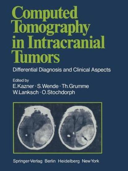
Computed Tomography in Intracranial Tumors

Computed Tomography in Intracranial Tumors

G.B. Bradac; U. Büll; R. Fahlbusch; Th. Grumme; E. Kazner; K. Kretzschmar; W. Lanksch; W. Meese; J. Schramm; H. Steinhoff; O. Stochdorph; S. Wende

Springer-Verlag Berlin and Heidelberg GmbH Co. K
2012
nidottu
The current book represents a distillation of the experience gained in diagnosis of intracranial tumors with computed X-ray tomography at the University Hos­ pitals of Berlin, Mainz, and Miinchen. To what purpose? Standard radiological techniques such as pneumoencephalography with lumbar puncture and cerebral arteriography with puncture of the common carotid artery are invasive proce­ dures which entail a certain amount of risk as well as discomfort for the patient. Furthermore, diagnoses made with these procedures rely primarily on indirect signs of an intracranial space-occupying lesion - such as displacement of the air-filled ventricles or of normal cerebral vessels. Only a few types of tumor are demonstrated directly with these techniques. In contrast, computed tomography demonstrates the pathology directly in almost all cases, and this with a minimum of risk and discomfort. In addition, normal intracranial structures are demonstrated, so that the tumor's effect on its surroundings can be evaluated. Today, almost a decade after HOUNSFIELD'S revolutionary invention, diagno­ sis of brain tumors without computed tomography is almost unthinkable, if not in fact irresponsible.
Advances and Technical Standards in Neurosurgery

Advances and Technical Standards in Neurosurgery

J. D. Pickard; C. Di Rocco; V. V. Dolenc; R. Fahlbusch; J. Lobo Antunes; M. Sindou; N. de Tribolet; C. A. F. Tulleken; M. Vapalahti

Springer Verlag GmbH
2003
sidottu
Epilepsy surgery is defined as any neurosurgical intervention whose pri- mary objective is to relieve medically intractable epilepsy (European Fed- eration of Neurological Societies Task Force 2000). The aim of epilepsy surgery is to reduce the number and intensity of seizures, minimise neuro- logical morbidity and antiepileptic drug (AED) toxicity, and improve quality of life. By definition, epilepsy surgery does not include normal surgical treatment of intracranial lesions where the primary goal is to di- agnose and possibly remove the pathological target, often an advancing tumour. In these patients, epileptic seizures are only one symptom of the lesion and will be treated concomitantly as part of the procedure. Temporal lobe epilepsy (TLE) is recognised as the most common type of refractory, focal epilepsy. In one third of all cases the neuronal systems responsible for the seizures that characterise this form of epilepsy fail to respond to currently available AEDs (Andermann F 2002). New imaging methods, especially magnetic resonance imaging (MRI), identify localising abnormalities in an increasing proportion of patients with intractable focal epilepsy. Consequently, the accuracy of the preoperative diagnostic pro- cedures has been significantly improved during the last decade; and suit- able candidates for surgery can be selected more reliably. Currently the main resources in most epilepsy surgery centres have been used to evaluate candidates for TLE surgery.