Kirjojen hintavertailu. Mukana 12 595 353 kirjaa ja 12 kauppaa.

Kirjailija

Surjith Vattoth

Kirjat ja teokset yhdessä paikassa: 3 kirjaa, julkaisuja vuosilta 2021-2025, suosituimpien joukossa Diagnostic Imaging: Head and Neck. Vertaile teosten hintoja ja tarkista saatavuus suomalaisista kirjakaupoista.

3 kirjaa

Kirjojen julkaisuhaarukka 2021-2025.

Diagnostic Imaging: Head and Neck

Diagnostic Imaging: Head and Neck

Bronwyn E. Hamilton; Bernadette L. Koch; Surjith Vattoth; Blair A. Winegar

Elsevier Health Sciences
2025
sidottu
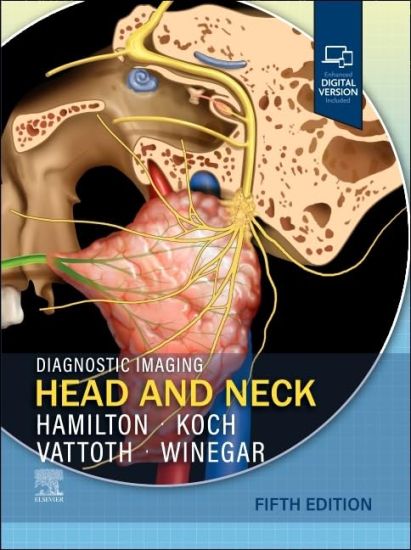
Covering the entire spectrum of this fast-changing field, Diagnostic Imaging: Head and Neck, Fifth Edition, is an invaluable resource for neuroradiologists, general radiologists, and trainees?anyone who requires an easily accessible, highly visual reference on today’s head and neck imaging. Drs. Bronwyn E. Hamilton, Bernadette L. Koch, Surjith Vattoth, Blair A. Winegar, and their team of highly regarded experts provide updated information on disease identification and imaging techniques to help you make informed decisions at the point of care. The text is lavishly illustrated, delineated, and referenced, making it a useful learning tool as well as a handy reference for daily practice. Serves as a one-stop resource for key concepts and information on head and neck imaging, including disease identification, imaging techniques, and details on tumor staging and classification Reflects recent updates in genetic and molecular characterization of tumors, congenital malformations, and inflammatory/autoimmune disorders, which all have implications for targeted precision therapies Offers more than 400 concise, informative chapters, dividing content into sections based on head and neck spaces and anatomic regions, with additional sections on cancers, posttreatment appearances, pediatric lesions, and syndromic diseases Features more than 2,800 high-quality print images (with an additional 3,800 images in the complimentary eBook), including radiologic images, full-color medical illustrations, clinical photographs, histologic images, and gross pathologic photographs Provides new and expanded content on genomic characterization of head and neck cancers; new characterizations of sinonasal tract and skull base tumors through distinctive genetic and molecular signatures (as reflected in the WHO’s 2022 reclassification); newer imaging techniques to characterize neuroendocrine tumors and spread using DOTATATE PET; whole-body imaging surveillance strategies for Li-Fraumeni syndrome, hereditary paraganglioma pheochromocytomas, and hereditary retinoblastoma; and the use of whole-body MR for tumor syndromes, multifocal vascular anomalies, and much more Emphasizes multidisciplinary involvement to help radiologists navigate the intersection of multiple specialties and confidently guide a wide variety of clinicians and surgeons to the appropriate diagnosis and treatment for a multitude of disorders Uses bulleted, succinct text and highly templated chapters for quick comprehension of essential information at the point of care Includes an eBook that allows you access to everything in the print version as well as additional images, text, and references, with the ability to search, customize your content, make notes and highlights, and have content read aloud; additional digital ancillary content may publish up to 6 weeks following the publication date
Imaging Anatomy: Head and Neck

Imaging Anatomy: Head and Neck

Surjith Vattoth

Elsevier Health Sciences
2024
sidottu
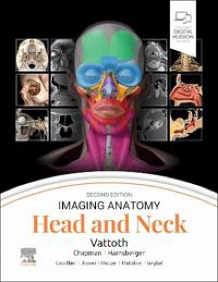
This richly illustrated and superbly organized text/atlas is an excellent point-of-care resource for practitioners at all levels of experience and training. Written by global leaders in the field, Imaging Anatomy: Head and Neck, second edition, provides a thorough understanding of the detailed normal anatomy that underlies contemporary imaging. This must-have reference employs a templated, highly formatted design; concise, bulleted text; and state-of-the-art images throughout that identify the clinical entities in each anatomic area, offering a unique opportunity to master the fundamentals of normal anatomy and accurately and efficiently recognize pathologic conditions. Features hundreds of detailed, full-color illustrations and more than 900 high-resolution, cross-sectional radiologic images that together illustrate the fine points of imaging anatomy for new and experienced head and neck imaging specialists Contains new chapters on external nose anatomy, the facial nerve in temporal bone, minor fissures and sutures around the temporal bone, and temporal bone anatomy on photon-counting detector (PCD) CT Provides updated, enlarged images and captions in areas such as facial muscles and the superficial musculoaponeurotic system, and frontal recess and related air cells Includes extensive new content on PCD CT; new details on relatively unknown anatomical foramina, such as the vomerovaginal canal and canaliculus innominatus; new content based on the International Frontal Sinus Anatomy Classification; and minute details on the course of nerves in the head and neck Includes a series of successive imaging slices in each standard plane of imaging (coronal, sagittal, and axial) to provide multiple views that further support learning Depicts common anatomic variants and covers the common pathological processes that manifest with alterations of normal anatomic landmarks Reflects new understandings of anatomy due to ongoing anatomic research as well as new, advanced imaging techniques Presents essential text in an easy-to-digest, bulleted format, enabling imaging specialists to find quick answers to anatomy questions encountered in daily practice Includes an eBook version that enables you to access all text, figures, and references with the ability to search, customize your content, make notes and highlights, and have content read aloud
Diagnostic Imaging: Head and Neck

Diagnostic Imaging: Head and Neck

Bernadette L. Koch; Surjith Vattoth; Philip R. Chapman

Elsevier - Health Sciences Division
2021
sidottu
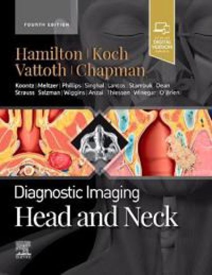
Covering the entire spectrum of this fast-changing field, Diagnostic Imaging: Head and Neck, fourth edition, is an invaluable resource for neuroradiologists, general radiologists, and trainees-anyone who requires an easily accessible, highly visual reference on today's head and neck imaging. Dr. Philip R. Chapman and his team of highly regarded experts provide up-to-date information on recent advances in disease identification, imaging techniques, and tumor staging to help you make informed decisions at the point of care. The text is lavishly illustrated, delineated, and referenced, making it a useful learning tool as well as a handy reference for daily practice. Serves as a one-stop resource for key concepts and information on head and neck imaging, including a wealth of new material and content updates throughout Features more than 2,800 illustrations including radiologic images, full-color illustrations, clinical and gross pathology photographs, and histology photographs, as well as an additional 2,200 digital images online Features numerous new chapters and updates from cover to cover including changes to staging of HPV-related/p16(+) oropharyngeal squamous cell carcinoma; new metastatic disease imaging recommendations, protocols, and treatments; and the latest knowledge on the genetics of various congenital conditions and syndromes Reflects new Lugano and WHO classifications for staging lymphomas; updates in the AJCC Cancer Staging Manual, 8th Edition; and updates from the 2018 ISSVA Classification regarding avoidance of outdated and inappropriate terminology and nomenclature that can lead to misdiagnosis or inappropriate treatments Uses bulleted, succinct text and highly templated chapters for quick comprehension of essential information at the point of care Enhanced eBook version included with purchase, which allows you to access all of the text, figures, and references from the book on a variety of devices