

Kirjailija
Torsten Bert Möller
Kirjat ja teokset yhdessä paikassa: 14 kirjaa, julkaisuja vuosilta 2008-2025, suosituimpien joukossa Taschenatlas Röntgenanatomie. Vertaile teosten hintoja ja tarkista saatavuus suomalaisista kirjakaupoista.
14 kirjaa
Kirjojen julkaisuhaarukka 2008-2025.


Pocket Atlas of Radiographic Anatomy
Torsten Bert Möller; Emil Reif
THIEME PUBLISHING GROUP
2025
nidottu
Radiographic anatomy at your fingertips. The Pocket Atlas of Radiographic Anatomy precisely describes the anatomical structures shown on conventional X-ray images and on the most important arthrographic and angiographic examinations. The double-page spread format makes the book extremely user-friendly: The X-ray image is directly juxtaposed with a detailed drawing showing the clinically relevant anatomical structures. This enables simple, clear, and rapid identification and naming of the anatomical structures shown on the X-ray images. Ideal for quick reference, the expanded 4th edition has been supplemented with new images, such as full-length views of the spine and leg, making it an indispensable resource for medical professionals and students alike.








Pocket Atlas of Sectional Anatomy, Volume III: Spine, Extremities, Joints
Torsten Bert Möller; Emil Reif
Thieme Publishing Group
2016
nidottu
Full multiplanar coverage of the spine, extremities, and joints! Renowned for its superb illustrations and highly practical information, the third volume of this classic reference reflects the very latest in state-of-the-art imaging technology. Together with Volumes 1 and 2, this compact and portable book provides a highly specialized navigational tool for clinicians seeking to master the ability to recognize anatomical structures and accurately interpret CT and MR images. Highlights of Volume 3: New CT and MR images of the highest qualityDidactic organization using two-page units, with radiographs on one page and full-color illustrations on the nextConcise, easy-to-read labeling on all figuresColor-coded, schematic diagrams that indicate the level of each sectionSectional enlargements for detailed classification of the anatomical structureComprehensive, compact, and portable, this popular book is ideal for use in both the classroom and clinical setting.

Pocket Atlas of Sectional Anatomy, Volume I: Head and Neck
Torsten Bert Möller; Emil Reif
Thieme Publishing Group
2013
pokkari
This comprehensive, easy-to-consult pocket atlas is renowned for its superb illustrations and ability to depict sectional anatomy in every plane. Together with its two companion volumes, it provides a highly specialized navigational tool for all clinicians who need to master radiologic anatomy and accurately interpret CT and MR images. Special features of Pocket Atlas of Sectional Anatomy:Didactic organization in two-page units, with high-quality radiographs on one side and brilliant, full-color diagrams on the other Hundreds of high-resolution CT and MR images made with the latest generation of scanners (e.g., 3T MRI, 64-slice CT) Consistent color coding, making it easy to identify similar structures across several slices Concise, easy-to-read labeling of all figuresUpdates for the 4th edition of Volume I: New cranial CT imaging sequences of the axial and coronal temporal bone Expanded MR section, with all new 3T MR images of the temporal lobe and hippocampus, basilar artery, cranial nerves, cavernous sinus, and more New arterial MR angiography sequences of the neck and additional larynx images Compact, easy-to-use, highly visual, and designed for quick recall, this book is ideal for use in both the clinical and study settings.
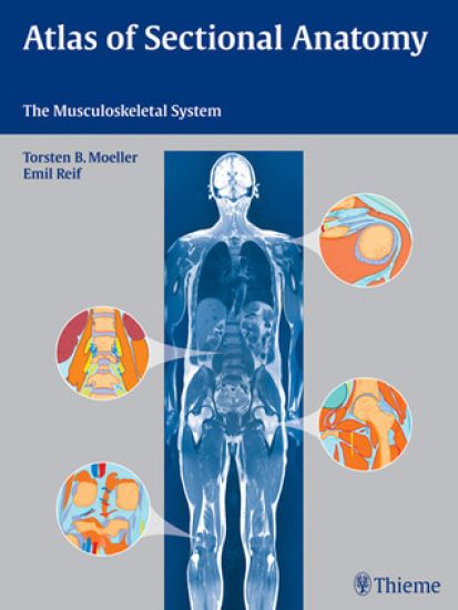
A handy, full-color resource for interpreting musculoskeletal MRI scans with confidence This superbly illustrated atlas provides a comprehensive presentation of the normal sectional anatomy of the musculoskeletal system to aid in the diagnosis of diseases affecting the joints, soft tissues, bones, and bone marrow. A precise, full-color drawing accompanies each high-quality sectional image, helping the reader to gain a solid understanding of the topographic anatomy and to differentiate between normal and pathologic conditions. Following examples of whole-body imaging, the atlas offers complete representations of the spinal column and the upper and lower extremities. The contiguous images of the extremities in transverse sections facilitate the identification of structures extending beyond the joints. Key features: Top-quality MRI scans, including whole-body views, produced with the most current, high-performance equipment Full-color illustrations drawn by the authors for optimal precision and accuracy Easy identification of anatomic structures through a uniform color code in the drawings Contiguous cross-sectional anatomy of the extremities Information on the location and direction of each slice for rapid orientation Atlas of Sectional Anatomy: The Musculoskeletal System is an invaluable reference for the daily practice of radiologists, radiology residents, and radiologic technologists.

Atlas der Schnittbildanatomie: Muskuloskelettales System
Torsten Bert Möller; Emil Reif
Georg Thieme Verlag
2008
sidottu